+86 17858656355
wang.fenglan@haolangmed.com
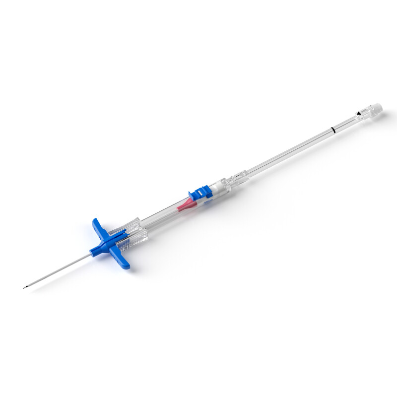
What is fibrin sheath on catheter?
By Zhao Qiaolin -Haolang Medical
It is the hot topic in clinical practice on how to prolong the catheter usage life. Studies have shown that fibrin sheaths form in almost all types of catheter access and the most common cause of catheter dysfunction is fibrin sheaths; In addition, it can also cause a series of complications, such as secondary infection, thrombosis, catheter removal, pulmonary embolism, etc. Therefore, early prevention, early detection and early intervention are necessary to avoid fibrin sheath formation which will leading to serious complications.
1.what is Fibrin sheath?
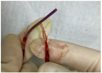
Images on the internet
Fibrin sheath is a proteinaceous film composed of endothelial cell, smooth muscle cell ,collagen and fibrous connective tissue, encasing the outer wall and endhole of the catheter, which lead to thrombosis, catheter dysfunction, secondary infection and hemal wall adhesion and decannulation difficulty, even complications such as pulmonary embolism formed after extubation. This kind of proteinaceous film has been successively described as fibrin sleeve, fibrin sheath, fibrin thrombus sleeve, sleeve and other names.
2.How does fibrin sheath occur?
At present, there are two viewpoints on the formation of fibrin sheath caused by catheter in domestic literatures of intravenous therapy.
The first view is that the fibrin sheath is a protein deposited in the blood on the surface of the catheter, formed by secondary thrombus, which is essentially a thrombosis.
The second view is that the formation of fibrin sheaths is a biological response of smooth muscle cells and endothelial cells in the vein wall to the catheter component and related thrombosis, it is a protective response of the vein wall to the catheter as a foreign body, and a process of self-regulation and repair of the body.
Studies have shown that this sheath only contains fibrin components in the early stage, but without fibrin components while mature, the thrombus with fibrin components are easy to fracture and can be dissolved by suppository, while mature sheath is dense fibrous connective tissue and cannot be dissolved by .Fibrin sheath is considered to be a structurally stable tissue. Some scholars observed the residual fibrin sheath in the blood vessels after extubation, it was still firmly attached to the venous wall 10 months after extubation.
3.The process of fibrin sheath formation
The endothelium of the vein wall is missing after catheter placing, then thrombosis is formed between the catheter and vein wall, becoming a thrombotic bridge. Gradually, the smooth muscle cells migrate from the venous wall through the thrombotic bridge to the catheter side, in addition, the endothelial cells of the venous wall will also crawl along the thrombotic bridge to the surface of the catheter. Finally, a sheath , consisting of smooth muscle cells and collagen as the main body, covered by endothelial cells ,is formed, and growing distal along the catheter wall.
4.The cause of formation of fibrin sheath
Relevant data shows that fibrin sheath formation is associated with catheter-associated thrombosis.
Three elements of thrombosis:
1. The injury of vascular endothelial
2, blood flow stasis
3, hypercoagulable state of the body
These three factors are the main causes of thrombosis. Studies have confirmed that catheter placement can directly cause vascular endothelial damage, and improper repeated puncture operation can also cause vascular endothelial damage. Besides,during the catheter indwelling, violent movement can lead to friction between catheter and vascular wall, causing mechanical stimulation of vascular wall and inducing thrombosis.In addition, elderly patients reduced activities after catheterization without enough drinking water, the catheter occupies the volume of blood vessels after catheterization, resulting in blood vessel narrowing and slow blood return. These elder patients’ blood is in a state of high coagulation, and they are the population with high incidence of thrombosis.The interaction of the above three points is the main cause of catheter-related thrombosis, and it is also considered by many experts and scholars to be the basis of the fibrin sheath.
5. When does the fibrin sheath form?
Some studies believed that the formation process of fibrin sheath is dynamic. There would be no fibrin sheath formed when catheter was placed within 6h. But,it start to be forming after 12h. The formed fibrin sheath has no clinical symptoms after the catheter indwelt for 1-2 weeks. The fibrin sheath in the later stages can cause catheter dysfunction and even difficulty in extubation.
Now accepted viewpoint is that the fibrin sheath begins to form at the point of contact between the catheter and the venous wall 24 hours after catheterization, and then extends along the catheter and gradually covers the entire catheter. It takes about 5-7 days to reach the full length of the catheter. Animal studies have confirmed that the incidence of fibrin sheath is 100% after 1 week of catheterization. Clinical confirmed that the incidence of fibrin sheath was 76% by angiography analysis. Some researchers also observed temporary hemodialysis catheters from catheterization to extubation by the color Doppler ultrasoundand for a month, found that almost all began to form mural thrombus at the place where the catheter entered the vein one day after catheterization, it is the beginning of the occurrence of fibrin sheath
6.The clinical manifestations signs of catheter-associated fibrin sheaths:
The fibrin sheath wraps around the surface of the catheter and may extend along the catheter, forming a live flap. Characteristic manifestations are the bolus from the catheter is unobstructed, while the live flap is attracted by negative pressure during ascent is blocked at the tip of the catheter, resulting in poor ejection and catheter dysfunction.
Common clinical manifestations of catheter disorders caused by fibrin sheaths are: exudate from the puncture point, slowing down the infusion rate, blockage of the catheter, no return of blood from catheter withdrawal, and difficulty extubation.
7. Imaging diagnosis of fibrin sheath
Currently, there is no standard for fibrin sheath imaging diagnosis. Some studies believe that the characteristic performance of color ultrasound imaging of fibrin sheath is that the catheter wall becomes thickening .Meanwhile, it is mentioned that color ultrasound has certain limitations, and ultrasound observation is not ideal for patients with thick fat layers.
8. How to treat the fibrin sheath?
1.Thrombolytic therapy with drugs, including urokinase streptokinase alteplase, which is pumped through a catheter into urokinase and contacts and dissolves fibrin erythrocytes, effectively prevents thrombosis and fibrous sheath formation.
2.In situ replacement of the dysfunctional or thrombus blocked cathete.
3.A stripping device can be placed in the vein to pull off the fibrin sheath from the top down, or a stripping device can be placed in the original catheter to break the fibrin sheath and remove it in situ. In this way, it is inevitable that the broken fibrin sheath will enter the blood circulation and enter the pulmonary artery, causing the possibility of pulmonary embolism.
9.Prevention of catheter fibrin sheaths
1. Standardized maintenance of the catheter after catheterization is the key to ensuring the normal use of the catheter and long-term retention.
2. Use vascular visualization technology to improve the success rate of puncture in patients with difficult intravenous access;
3. According to the patient's vascular conditions and treatment needs, select the appropriate catheter model and blood vessel;
4. The vortex generated by the push-stop-push flushing method can effectively wash away the attachments and drugs attached to the inner wall of the catheter and the positive pressure sealing method, which can not only reduce the occurrence of catheter blockage, but effectively prevent the formation of fibrin.
5. Select a 10ml syringe to rinse the fibrin tail or fibrin sheath at the tip of the catheter by accelerating the frequency of flushing tube, because the smaller the size of the syringe, the greater the pressure of the syringe, there is a possibility of damage to the catheter, vascular endothelium, and the larger size of the syringe may not achieve the flushing effect.
6. If blood is drawn, transfused, or other viscous fluids are drawn through this catheter, the tube must be flushed manually before other fluids are injected.
7.Frequently observe the catheter drip rate, and when the drip rate is found to slow down, the cause should be found out in time and properly handled.
8. Frequently observe whether there are special circumstances such as redness, swelling, induration, exudate and so on at the insertion point, and local treatment should be done in time.
9. Patients are instructed to wear loose-sleeved clothing, because too tight sleeves will hinder blood circulation and accelerate thrombosis.
10. Strengthen home education for outpatients, inform patients to closely observe the arm circumference of both upper limbs, measure the arm circumference once a day, and make a record.
11. Encourage the use of various non-drug methods to prevent blood clots, early movement of the limb, mild exercise, and drinking enough water.