+86 17858656355
wang.fenglan@haolangmed.com
In recent years, the incidence of critically ill neonates has increased significantly. After admission to the Neonatal Intensive Care Unit (NICU), these infants often require the placement of intravenous catheters for the administration of medications and nutritional support. Clinical guidelines recommend the preferential use of central venous access when infusing irritant agents such as calcium preparations or vasoactive drugs. However, in emergency resuscitation scenarios or when central venous catheterization fails, peripheral intravenous catheter placement is a commonly adopted alternative.
Due to their small vessel diameter, fragile vascular walls, limited puncture sites, and low cannulation success rates, neonates are at a high risk of extravasation—where infused solutions leak into the surrounding tissues—during peripheral intravenous therapy. Studies have shown that the incidence of extravasation in NICU patients is 11.7%. Mild extravasation may cause localized erythema, swelling, and pain, whereas severe extravasation can lead to tissue necrosis, limb dysfunction, and even adverse outcomes that may affect the infant’s prognosis.
This study systematically searched and reviewed both domestic and international literature on the prevention and management of peripheral intravenous extravasation in NICU infants. Twenty-five evidence-based recommendations were extracted, integrated, and summarized from six key domains: risk factors, catheter placement, catheter maintenance, extravasation assessment, extravasation management, and team building with education and training. These findings provide a clinical reference to guide practice. Healthcare professionals are advised to carefully select and apply evidence-based strategies in accordance with the clinical context and the infant’s specific condition to ensure the safety of peripheral intravenous therapy.
Table 4 Summary of the Best Evidence for Prevention and Management of Extravasation in NICU Neonates
|
Evidence Theme |
Evidence Content |
Evidence Grade (Level) |
|
Risk Factors |
1. Patient factors: Unstable circulation, extremely low birth weight, post - 3/4 - level surgery state, infection, presence of diseases that may cause blood vessel or circulation damage (such as diabetes, lymphatic edema, neonatal asphyxia, perineural lesions, perivascular diseases) [2,6,15,17] |
3b |
|
2. Puncture and catheter factors: Multiple punctures leading to vascular sclerosis, catheter tip located near a joint, use of butterfly - type steel needle infusion devices, improper catheter fixation, catheter indwelling time > 24 h [2,3,14,17] |
3b |
|
|
3. Drug factors: Infusion of fluids with osmolality > 900 mOsmol/L, pH < 5 or > 9, large - volume rapid drug infusion (such as high - pressure injection, high - speed infusion) [2,3,14,17] |
4a |
|
|
Catheter Insertion |
4. Before the first catheter insertion, identify extravasation risk factors. Based on the expected treatment duration, drug characteristics, and available peripheral vein puncture sites, plan the selection of peripheral veins and puncture sites rationally [2,6,12,15] |
4a |
|
5. Follow the general order of choosing veins on the back of the hand, foot, scalp, and cubital fossa, and select visible, palpable, and elastic veins in sequence for catheter insertion [2,6,15] |
1b |
|
|
6. When puncture is difficult, methods such as local application of warm compresses, tapping, and massage can be used to dilate blood vessels, or near - infrared spectroscopy can be used to assist puncture; when 2 people fail to puncture 2 times, consider seeking help from a Intravenous Therapy Team in a timely manner [2] |
1a |
|
|
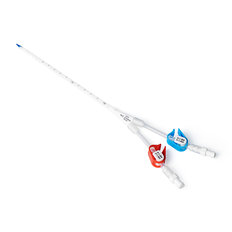
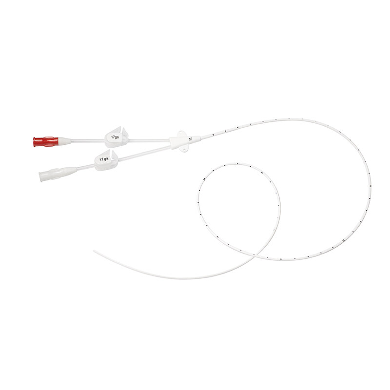
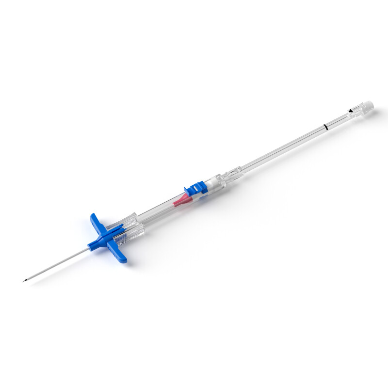
7. Under the condition of meeting the treatment needs, preferentially select the smallest - gauge, silicone or polyurethane catheters. For neonates, catheters of 22 - 26 G can be used [2,16] |
1b |
|
|
8. At least 2/3 of the catheter length should be within the blood vessel. When fixing, use sterile materials to cover the entire catheter with the puncture point as the center. Do not affect the observation of the puncture point and the speed of Intravenous Infusion [2,6,17] |
1b |
|
|
Catheter Maintenance |
9. Use non - drug - based interventions such as restraint to reduce neonatal movement, improve the success rate of the first puncture, and facilitate catheter fixation [2,6,20] |
1b |
|
10. When the risk of catheter displacement due to the use of topical anesthetics is high, a catheter fixation device (puncture site protection or physical fixation device) can be used, but it should not affect the observation of the puncture site and the vascular access. The dressing should be removed as soon as possible [2,7] |
2b |
|
|
11. Use a disposable non - negative pressure tube flushing solution for pulse - style flushing. The flushing volume should be based on the drugs in the catheter and the lumen of the additional device. After flushing, it should be flushed and pressurized to twice the lumen volume of the catheter and the additional device [2,6,7] |
1a |
|
|
12. According to the situation, the positive pressure tube sealing solution should be at least the sum of the lumen volumes of the catheter and the additional device, and the tube sealing volume should be 1.2 times the sum of the lumen volumes of the catheter and the additional device [2,7] |
2b |
|
|
13. Before infusing drugs, flush the tube to confirm that the catheter is in the vein; after infusing hypertonic solutions, antibiotics, vasoactive drugs, etc., which have relatively large vascular irritation, immediately perform pulse - style flushing; for peripheral veins that are not in use but must be retained, flush and seal the tube at least every 24 h [2,7] |
1b |
|
|
14. When infusing parenteral nutrition solutions outside the 900 mL/week limit, the glucose concentration should be restricted to ≤ 10.0%, the protein concentration ≤ 5%, and the osmolality ≤ 900 mOsmol/L [2,3,15] |
2a |
|
|
Extravasation Assessment |
15. Assess whether extravasation occurs by observing for redness or pale skin around the puncture point, serous exudation, local temperature increase, and the presence of crying, painful expressions and other pain manifestations in the patient, as well as swelling at the infusion site, and comparing the circumference of the bilateral limbs. Do not rely on the infusion pump alarm to identify extravasation [2,3,6,14,17] |
5b |
|
16. Assess daily during intermittent infusion and hourly during continuous infusion; assess when changing dressings before and after each infusion; when there are extravasation risk factors, increase the assessment frequency and conduct dynamic assessment [2,17] |
1b |
|
|
17. When extravasation occurs, assess and record the injury degree: Stage Ⅰ (local tissue inflammatory reaction stage), local skin redness, swelling, heat, pain, no blisters; Stage Ⅱ (phlebitis stage), local subcutaneous tissue bleeding or blister formation, blisters rupture, tissue pale, forming a superficial ulcer; Stage Ⅲ (tissue necrosis stage), local skin gangrene, combined with black scab, deep ulcer, muscle, blood vessel, nerve exposure or infection [2] |
5b |
|
|
Extravasation Management (Non - pharmacological Measures) |
18. When the extravasated fluid is an acidic or alkaline drug, non - pharmacological measures should be used [2] |
2b |
|
19. Immediately stop the infusion, try to aspirate the extravasated drug as much as possible, remove the catheter after aspiration; raise the affected limb by 20° - 30°, and avoid pressure on the extravasation site [2,3,18] |
2b |
|
|
20. Within 24 - 48 h after extravasation, physical cooling can be used for drugs that cause extravasation and are heat - dissipating; for drugs that cause extravasation and are heat - diffusing, cold compresses (temperature 4 - 6 °C) can be used. The time of cold/hot compresses should be ≤ 42 °C [2,17] |
5a |
|
|
21. When the blister diameter caused by extravasation is > 0.5 cm, it is recommended to aspirate the blister fluid under sterile conditions when the blister tension decreases, and cover with sterile dressings; when extravasation causes tissue necrosis, the necrotic tissue should be debrided immediately by personnel who have received relevant training, debrided every 2 d, and skin grafting should be performed if necessary [2,17] |
5b |
|
|
Pharmacological Management Measures |
22. When the extravasated fluid is a high - concentration electrolyte (sodium, potassium, calcium) solution, parenteral nutrition solution, antibiotic, sodium bicarbonate, or a drug with an osmolality > 800 mOsmol/L, or when there is minimal subcutaneous fat at the extravasation site, hyaluronidase should be injected subcutaneously into the four quadrants around the puncture site for local infiltration. Injection within 1 h of extravasation is more effective, and it can be used within 12 h. Hot compresses can be used concomitantly [2,17,18]. |
3b |
|
23. When the extravasated fluid is a vasopressor, phentolamine should be injected subcutaneously into the four quadrants around the puncture site. Injection within 1 h of extravasation is more effective, and it can be used within 12 h. Alternatively, 2% nitroglycerin can be applied topically 2–3 cm around the extravasation site, and repeated every 8 h if necessary. Closely monitor for adverse drug reactions during use, including hypotension, tachycardia, and arrhythmia, especially in premature infants, with simultaneous blood pressure monitoring [2,17,18]. |
1b |
|
|
Team Building, Education, and Training |
24. Establish a pediatric intravenous therapy team. Team members should include pediatric or neonatal specialized nurses and intravenous therapy specialized nurses, all of whom should hold municipal - level or higher qualifications. Team members should regularly analyze harmful extravasation incidents and implement quality improvement measures [2,6]. |
3b |
|
25. Neonatal nurses should receive training on peripheral intravenous catheter insertion and maintenance, including professional theoretical knowledge and skills training. Regular assessments should be conducted to evaluate nurses’ understanding of relevant guidelines and their implementation in practice [2,7,19]. |
1c |