+86 17858656355
wang.fenglan@haolangmed.com
In clinical practice, peripherally inserted central catheters (PICCs) are most commonly placed via the upper-extremity veins. However, in certain special situations, upper-limb access may be unfeasible or carry elevated risks. In such cases, lower-extremity PICC placement can serve as an important alternative. This article presents a successful case of lower-extremity PICC insertion and discusses key technical considerations, nursing strategies, and clinical implications.
Case Background
The patient was an 82-year-old male with a chronic debilitating condition requiring prolonged hospitalization and intravenous therapy. Following the current admission, his clinical status further deteriorated, necessitating continuous infusion of vasoactive medications and ventilatory support. The upper-extremity venous access was extremely compromised due to a history of multiple previous catheterizations, venous stenosis, and severe edema. He had developed PICC-related venous thrombosis in the right upper limb, and there was a prior history of thrombosis in the left lower limb.
Traditional upper-extremity PICC placement was therefore not feasible. However, the patient required reliable long-term venous access for continued treatment, and the family requested active management. After multidisciplinary evaluation by the medical team and the vascular access nursing specialists, lower-extremity PICC placement was considered and ultimately selected as the intervention.
Overcoming Challenges
Clinical experience with lower-extremity PICC placement is relatively limited. This patient was bedridden long-term, unable to mobilize independently, and had a history of thrombosis in the left lower limb—factors that significantly increased the risk of post-procedure thrombosis. To ensure a successful catheter placement, the clinical team conducted extensive preparatory work, reviewed literature and technical guidelines on lower-extremity PICC insertion, developed contingency plans, and thoroughly communicated with the family to explain the necessity and potential risks prior to proceeding with cannulation.
Challenge 1
For lower-extremity PICC placement, the femoral vein is typically the first-choice access site. However, the femoral vein lies in close proximity to the femoral artery and femoral nerve, increasing the risk of inadvertent arterial puncture or nerve injury.
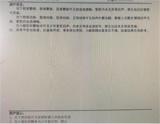
This patient had a thigh circumference of 54 cm. Ultrasound examination showed that the femoral vein was located beneath the femoral artery along most of its course. Only a single point in the mid-thigh demonstrated a slight separation of the artery and vein, where the femoral vein was positioned at the 7 o’clock direction relative to the artery at a depth of 2.5 cm. This anatomical relationship greatly increased the difficulty of puncture.
With no alternative access options available, the team proceeded despite the challenges.
Challenge 2
After identifying the puncture site, optimal patient cooperation was still needed to maintain adequate external rotation and stabilization of the thigh. However, the patient’s prolonged bed confinement had resulted in partial muscle atrophy, reduced joint mobility, and limited positioning capacity. To facilitate the procedure, a pillow was placed beneath the thigh to elevate and support the limb.
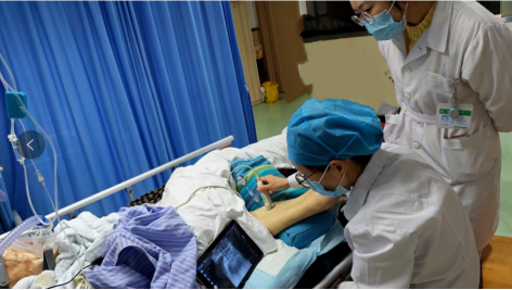
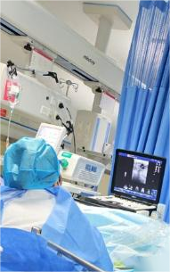
Under ultrasound guidance, the puncture needle was advanced gradually. The needle successfully bypassed the femoral artery and entered the femoral vein, but no blood return was observed. The ultrasound probe was then rotated into the longitudinal axis, clearly visualizing the needle tip within the lumen. With assistance, the guidewire was advanced, and ultrasound confirmed its smooth entry into the vessel—indicating successful venous cannulation.
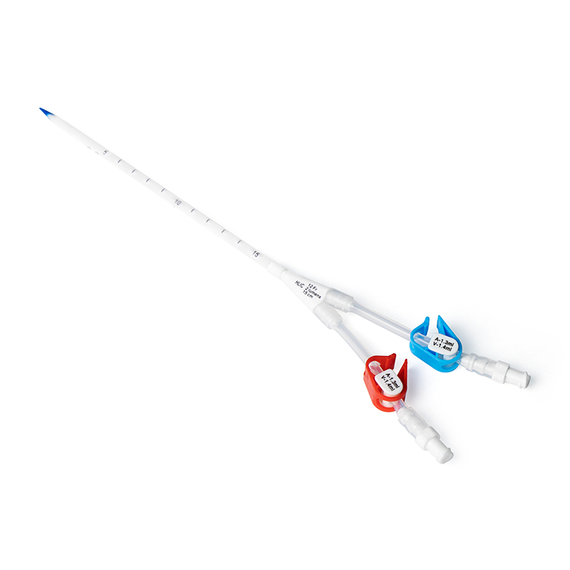
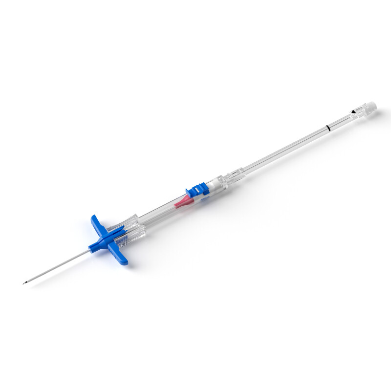
Following catheter placement, an abdominal X-ray confirmed that the catheter shadow was visible in the right iliac vein and inferior vena cava, with the distal tip located at the level of the L2 vertebral body. After a challenging one-hour procedure, a critical lifeline was successfully established for the patient.
Safeguarding the Lifeline
Lower-extremity PICC placement is suitable for patients with poor upper-extremity venous access, superior vena cava syndrome, or special positioning requirements. A thorough evaluation of lower-limb venous status and mobility is essential prior to the procedure.
Post-insertion care is equally critical. Monitoring the puncture site, measuring thigh circumference, performing routine catheter maintenance, and preventing infection and venous thrombosis all play vital roles in protecting this lifeline.
By breaking away from traditional approaches and adopting an alternative route, lower-extremity PICC placement provides an important venous access option for patients with special needs. With precise technique and standardized nursing care, lower-extremity PICC insertion can be performed safely and effectively to meet clinical requirements, offering new insights and reference strategies for managing similar patients.