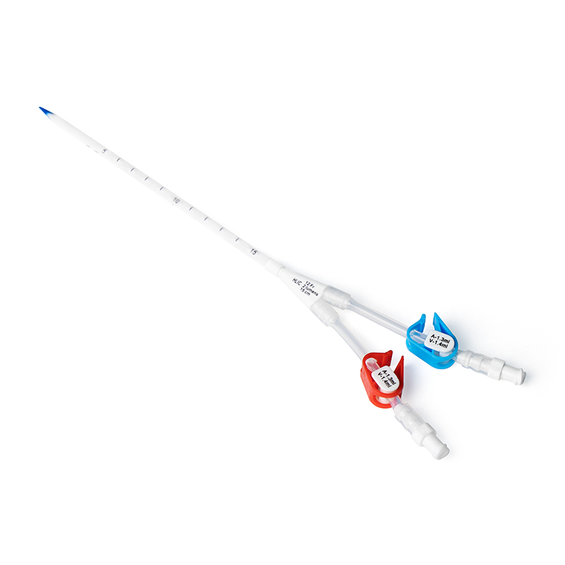
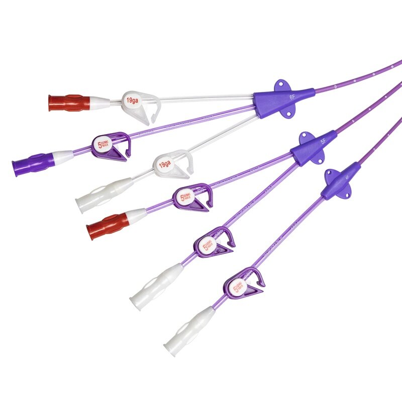
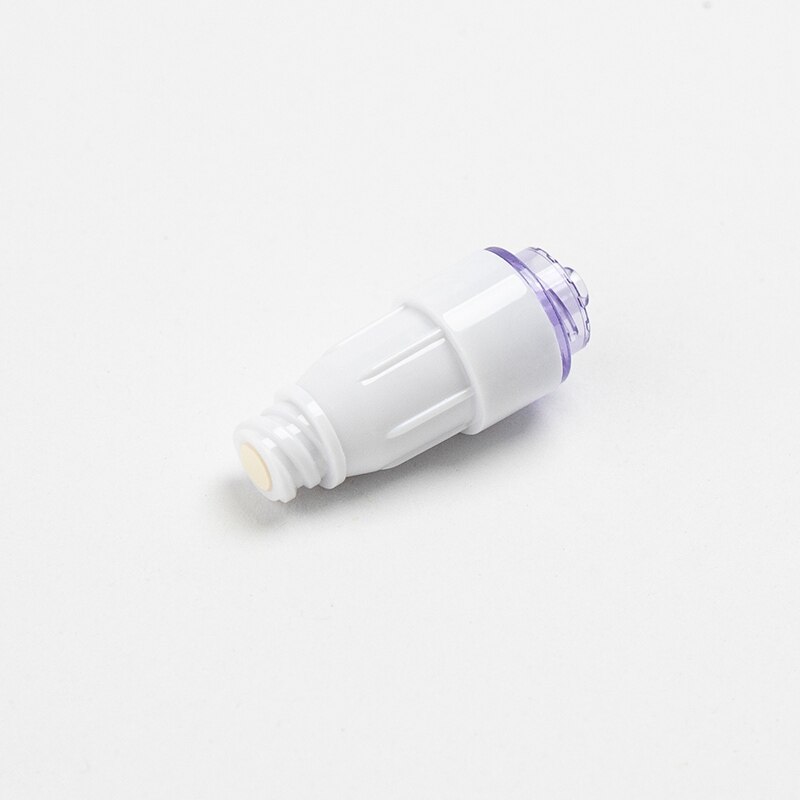
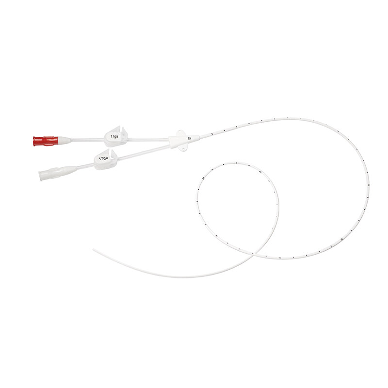
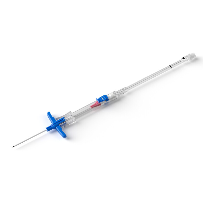
The 9th edition of the Infusion Therapy Standards of Practice places greater emphasis, compared with the 8th edition, on the importance of carefully assessing and selecting the most appropriate vascular access device (VAD) and insertion site, as well as verifying device tip location and patency before and during infusion therapy, in order to reduce the risk of infiltration and extravasation. The updated standards also include more detailed recommendations regarding risk reduction strategies, assessment of related risk factors, recognition of signs and symptoms, and appropriate interventions.
Strategies to Reduce the Risk of Infiltration/Extravasation
1. For patients with difficult venous access, it is recommended to escalate early to a vascular access specialist or specialized team.
2. Strengthen staff training on risk factors for infiltration/extravasation, optimal site selection for VAD placement, and recognition and management strategies.
3. Implement insertion and management of peripheral intravenous catheters in accordance with clinical practice guidelines, as this can reduce extravasation rates.
4. Establish a case repository of extravasation events to support education and training.
5. Identify optimal VAD insertion sites for contrast media administration, and assess device location and local conditions before, during, and after infusion of contrast media.
Risk Factors Associated with Infiltration/Extravasation
1. Patient-related risk factors: communication barriers in infants and young children; fragile skin and veins; age-related deterioration of skin and vascular structures in older adults; gradual malposition of long-term central venous access devices in infants or children as they grow.
2. Peripheral IV catheter–related factors: use of steel-winged “butterfly” needles; inadequate catheter securement; peripheral IV catheters placed in the hand, wrist, foot, ankle, antecubital fossa, or other sites with minimal subcutaneous tissue coverage.
3. Mechanical risk factors: abnormalities such as fibrin sheath formation, venous thrombosis, pinch-off syndrome, or catheter fracture, which may result in device and/or vessel occlusion; vascular trauma caused by rapid, large-volume infusions, frequent cannulation of the antecubital fossa, or the use of large-bore catheters.
Recognition of Signs and Symptoms of Infiltration/Extravasation
1. Promptly identify pain, paresthesia, or circulatory impairment following VAD insertion in small-caliber veins or at joint sites.
2. Observe for discoloration or hyperpigmentation in areas proximal and distal to the insertion site.
3. Rule out other conditions presenting with similar symptoms, such as phlebitis, flushing reactions, or rash.
Interventions for Infiltration/Extravasation
1. Once infiltration or extravasation is detected, immediately stop the infusion. Elevate the affected limb to the level of the heart, if appropriate, to promote venous return (note: elevation is contraindicated in patients with compartment syndrome).
2. Select treatment strategies based on the patient’s symptoms, as well as the type and volume of solution/medication infiltrated into the surrounding tissues. Appropriate antidotes should be administered when indicated, for example: sodium thiosulfate for extravasation of mechlorethamine, bendamustine, calcium, or cisplatin. Avoid cold compresses in cases of vasoconstrictor extravasation. For contrast media extravasation, if inflammation peaks within 24–48 hours:
For extravasation volumes <50 mL, conservative management and cold compresses are recommended.
For volumes >50 mL accompanied by circulatory or neurologic impairment, immediate surgical consultation is required.
3. For severe tissue injury due to extravasation, treatment options may include negative-pressure wound therapy, aspiration, dressings with ethacridine lactate and phototherapy, decellularized fish-skin grafts, dehydrated human amniotic membrane allografts, and laser therapy to reduce skin discoloration caused by hemosiderin deposition.
4. Provide outpatient or telephone follow-up to assess the progression of the extravasation.
References
1. Indarwati F, Mathew S, Munday J, et al. Incidence of peripheral intravenous catheter failure and complications in paediatric patients: systematic review and meta-analysis. International Journal of Nursing Studies. 2020;102:103488.
2. Karaoğlan N, Sarı HY, Devrim İ. Complications of peripheral intravenous catheters and risk factors for infiltration and phlebitis in children. British Journal of Nursing. 2022;31(8)\:S14–S23.
3. Melo JMA, Oliveira PP, Souza RS, et al. Prevention and conduct against the extravasation of antineoplastic chemotherapy: a scoping review. Revista Brasileira de Enfermagem. 2020;73(4)\:e20190008.
4. Taibi A, Bardet MS, Durand Fontanier S, et al. Managing chemotherapy extravasation in totally implantable central venous access: use of subcutaneous wash-out technique. The Journal of Vascular Access. 2020;21(5):723–731.
5. Marsh N, Webster J, Ullman AJ, et al. Peripheral intravenous catheter non-infectious complications in adults: a systematic review and meta-analysis. Journal of Advanced Nursing. 2020;76(12):3346–3362.
6. Kim JT, Park JY, Lee HJ, et al. Guidelines for the management of extravasation. Journal of Educational Evaluation for Health Professions. 2020;17:21.
7. Massand S, Carr L, Schneider E, et al. Management of intravenous infiltration injuries. Annals of Plastic Surgery. 2019;83(6)\:e55–e58.
8. Ren P, Cao J, Gao P, et al. Interpretation of vascular access device complications in the 2024 Infusion Therapy Standards of Practice by the Infusion Nurses Society. Chinese Journal of Nursing Research. 2025;39(15):2497–2503.

